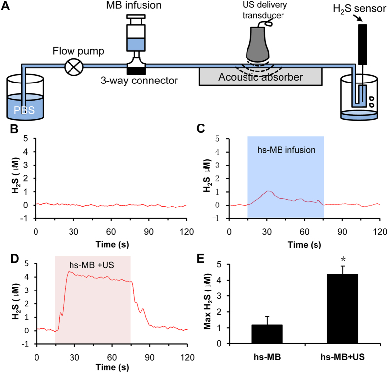
Figure 2. Ultrasound triggered H2S release from hs-MB in vitro.
(A) In vitro setup of flow system for ultrasound triggered H2S release from hs-MB. (B) Baseline level of H2S. (C) Change of H2S level during hs-MB infusion. (D) Change of H2S level during hs-MB infusion and ultrasound irradiation. (E) Comparison of maximum concentration of H2S. *P < 0.05, vs hs-MB. US indicated ultrasound; hs-MB, microbubble loaded with hydrogen sulfide.

