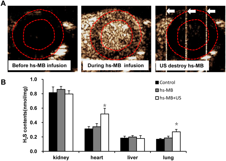
Figure 3. In vivo local delivery of H2S mediated by hs-MB and US.
(A) In vivo imaging of ultrasound targeted hs-MB destruction in the myocardium. Red dotted line indicated the region of myocardium. White arrow indicated the ultrasound pulse emitted from an ultrasonic cavitation apparatus. (B) Comparison of H2S concentration in various tissues following treatment. *P < 0.05 vs Control. US indicated ultrasound; hs-MB, microbubble loaded with hydrogen sulfide.

