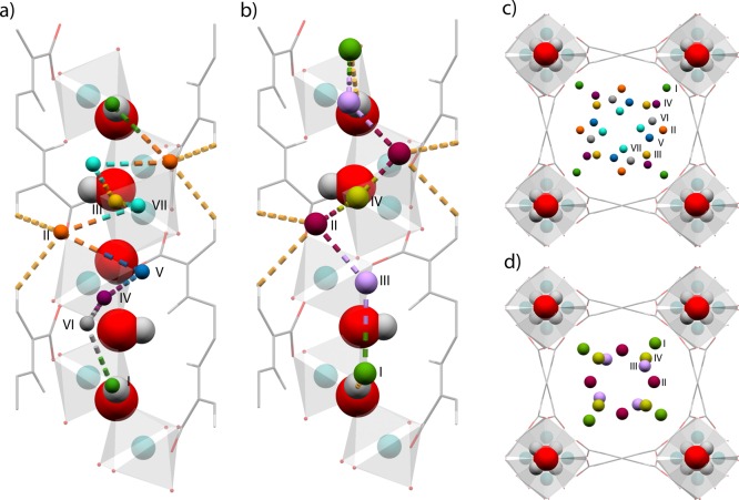
Figure 2.
View of the crystal structure of H2/CH4 loaded MFM-300(In) showing (a,b) view of the corner-sharing extended [InO4(OH)2] chain highlighting interactions between the framework structure with D2 and CD4 molecules, respectively; and (c,d) view of the c-crystallographic axis showing the positions of the adsorbed D2 and CD4 molecules, respectively. D2 molecules in (a) and (c) are colored according to the scheme: Site I, green; Site II, orange; Site III, yellow; Site IV, purple; Site V, blue; Site VI, gray; Site VII, light blue. CD4 molecules in (b) and (d) are colored according to the following scheme: Site I, green; Site II, orange; Site III, yellow; Site IV, purple; Site V, blue; Site VI, gray; Site VII, light blue.

