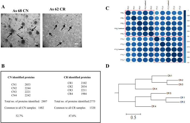
Figure 1. Morphological features of ascites-derived spheroids.
(A) Representative light microscopy images of non-adherent spheroid tumor populations grown on low-attachment plates derived from CN and CR serous ovarian cancer patient ascites samples. Images are representative of n = 4 samples/group. Arrows indicate non-adherent spheroid ascites tumour populations. Magnification is set at 100x, scale bar = 100μM. (B) CN and CR non-adherent spheroid cellular samples were profiled using discovery-based proteomics. Samples were analyzed as technical replicates (n = 2), stringent peptide and protein identification criteria were implemented (1% FDR protein, 5% PEP), with proteins requiring at least two significant peptides for identification. For CN samples (2807 proteins identified, 1482 proteins in common across CN1-4), while for CR samples (2775 proteins identified, 1320 proteins in common across CR1–4). (C) Correlation matrix of CN (1–4 replicates and combined) and CR (1–4 replicates and combined) samples showing that each individual sample represents high similarity with other sample replicates of the same cohort. (D) Cluster analysis of CN and CR replicates showing clear distribution between sample groups.

