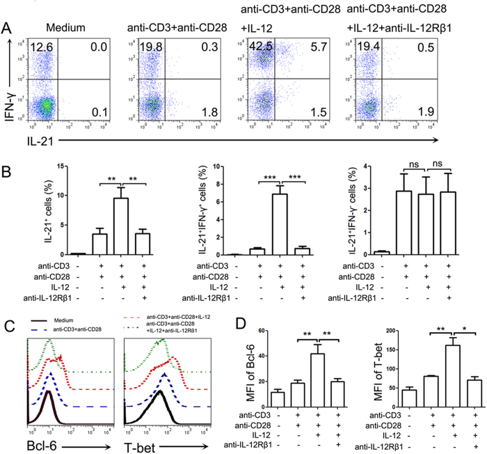
Figure 6. IL-12 directly regulated purified CD8+ T cells to express IL-21 and IFN-γ.
Purified CD8+ T cells from PBMCs of NP patients were stimulated with anti-CD3 and anti-CD28 in the presence or absence of IL-12 and anti-IL-12Rβ1 antibodies for 72 h. PMA and ionomycin plus BFA were added in the last 5 h, and the cells were assayed with FACS. (A) Representative data show the IL-21 and IFN-γ expression levels by CD8+ T cells. (B) The statistical results show the percentage of IL-21+, IL-21+IFN-γ+ and IL-21+IFN-γ− cells (n = 18) amongst the CD8+ T cells. (C,D) The representative FACS data and summary results show the Bcl-6 and T-bet expression levels by the CD8+ T cells (n = 18). *P < 0.05; **P < 0.01; ***P < 0.001; ns, no significance.

