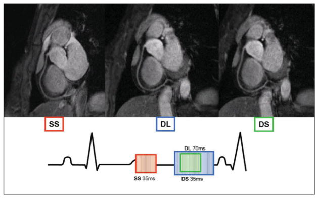
Figure 1.
Coronary MRA of the RCA obtained in the same subject in diastole using a long (blue frame) and short (green frame) acquisition window. Image acquired using a short acquisition window at end-systole (red frame). Same color scheme is used on the electrocardiogram tracing to indicate position of acquisition in the RR interval. DL = diastolic and long acquisition window, DS = diastolic and short acquisition window, SS = systolic and short acquisition window.

