Figure 1.

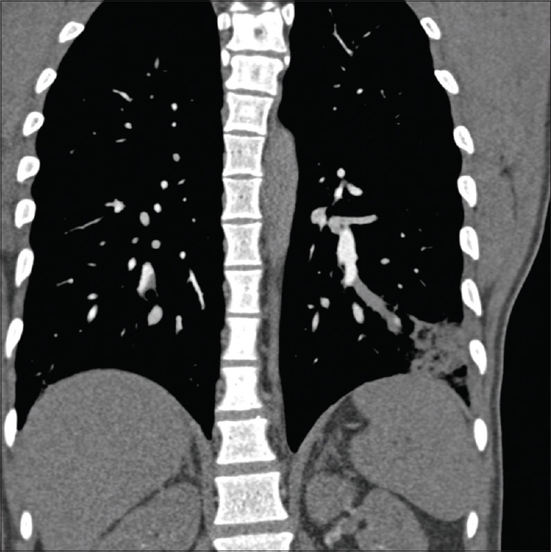
Computerized tomography pulmonary angiography showing filling defect in segmental branch supplying lateral basal segment of left lower lobe along with consolidation of corresponding lung segment suggestive of pulmonary embolism

Computerized tomography pulmonary angiography showing filling defect in segmental branch supplying lateral basal segment of left lower lobe along with consolidation of corresponding lung segment suggestive of pulmonary embolism