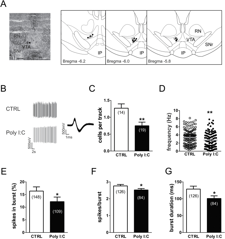
Figure 4.
Prenatal polyriboinosinic-polyribocytidilic acid [poly(I:C)] treatment dysregulates dopamine neuron firing activity in adulthood. (A) Example of a recording location for a ventral tegmental area (VTA) dopamine neuron in a poly(I:C)-treated rat (the triangle indicates the pontamine sky blue dye). The diagrams at right show samples of localizations of recording sites in poly(I:C) rats (black dots) and controls (white dots) as verified by histological sections. IP, interpeduncular nucleus; RN, red nucleus; SNr, substantia nigra pars reticulata. Scale bar = 0.5mm. (B) This panel shows traces illustrating representative extracellular recordings of a putative dopamine neuron in the VTA of anesthetized rats belonging to the control group (CTRL, above) and poly(I:C) group (below). Dopamine neurons recorded from poly(I:C) rats typically show slower firing activity and a reduced bursting compared with CTRL. The left trace shows the typical broad spike waveform of a dopamine neuron. (C) The bar graph shows the number of spontaneously active VTA dopamine neurons, which was different between the experimental groups. The scatter plot in (D) displays individual dopamine neuron firing rate in CTRL and poly(I:C) rats. The horizontal lines represent average values that are significantly different between the 2 groups. Graph histograms represent the percentage of spikes in bursts (E), the mean number of spikes per each burst (F), and the mean burst duration (G). Data are expressed as percentage or mean±SEM. *P<.05, **P<.01.

