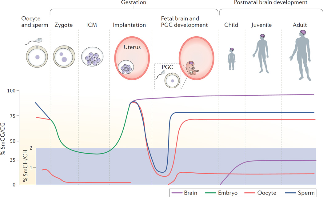
Figure 3. DNA methylation landscape across development.
DNA methylation for 5-methylcytosine-guanine (5mCG) and 5-methylcytosine-(A, T or C) (5mCH) is shown on separate scales across human development. 5mCG levels in mature germ cells (sperm and oocyte) decrease after fertilization (zygote) and continue to decline in the inner cell mass (ICM) until implantation, when they rise rapidly. Primordial germ cells (PGCs) in the developing fetus then undergo a second global demethylation and subsequent re-methylation during maturation. Somatic cells, including brain cells, gradually gain 5mCG during the remainder of fetal and postnatal development. 5mCH levels are detectable in the oocyte (but not sperm), decline post-fertilization and return during subsequent PGC development. In the brain, 5mCH is detectable in glia and occurs at relatively high levels in neurons compared with other cell types. 5mCH levels in neurons rise after birth, peak in early childhood and are maintained into adulthood.

