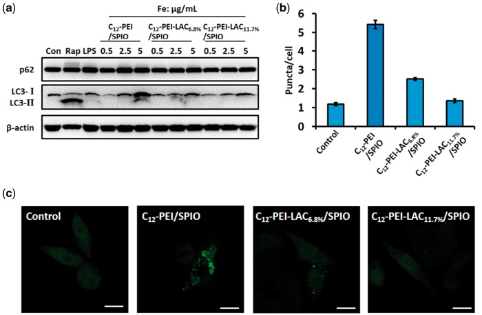
Figure 3.
C12-PEI/SPIO nanocomposites induced autophagy can be prevented by PEI lactosylation. (a) Western blotting assay of RAW 264.7 cells that were untreated (negative control), treated with rapamycin (50 μg/ml, 8 h), LPS (1 μg/ml 24 h) and nanocomposites (0.5, 2.5, 5 μg/ml, 24 h). Both rapamycin and LPS groups served as positive controls. The RAW 264.7 cells stably expressing GFP-LC3 were labelled with nanocomposites at an iron concentration of 5 μg/ml for 24 h. (b) the GFP puncta in RAW 264.7 cells were measured and summarized. Data were the mean value of three independent experiments with each count of no less than 60 cells. Values are expressed as the mean ± SD, n = 3. (c) the representative images of GFP puncta in RAW 264.7 cells after treatment under a CLSM. Scale bar: 10 μm.

