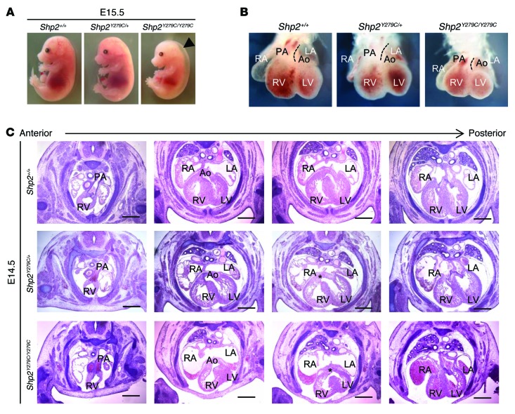
Figure 2. NSML embryo hearts at E14.5 display alignment and VSDs, leading to partial embryonic lethality.
(A) Representative photomicrographs of E15.5 littermate embryos from heterozygous Shp2Y279C/+ mating. (B) Bright-field whole-mount frontal view of hearts from E14.5 littermate embryos. Dotted lines delineate the leftmost border of the pulmonary artery, highlighting the relative leftward shift of the pulmonary artery and corresponding rightward shift of aorta in Shp2Y279C/Y279C embryo hearts. (C) Cardiac morphology of anterior to posterior H&E-stained heart sections of littermate embryos. Note, both aorta and pulmonary artery originate from the right ventricle in the Shp2Y279C/Y279C embryo (second column, lower panel). In addition, both Shp2Y279C/+ and Shp2Y279C/Y279C embryos show VSDs, as indicated by an asterisk (third column, middle and lower panels). Scale bars: 400 μm. PA, pulmonary artery; RV, right ventricle; LV, left ventricle; Ao, aorta; LA, left atria; RA, right atria.

