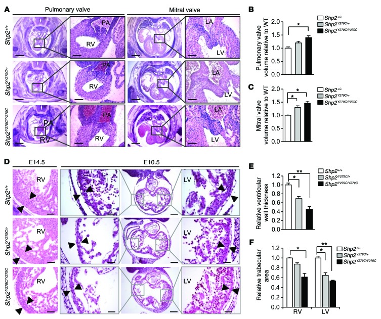
Figure 3. NSML embryo hearts display enlarged endocardial cushions, decreased trabeculation, and a thinner myocardial wall.
(A) H&E staining of endocardial cushions of developing pulmonary and mitral valve leaflets at E14.5. Scale bars: 400 μm; 100 μm (inset). Quantification of (B) pulmonary and (C) mitral valve volumes of NSML hearts relative to control (Shp2+/+). n = 3 embryos/group. (D) H&E staining of ventricular wall at E14.5 and E10.5, respectively. Note the thinning of the compact zone (black arrowheads) and a decreased trabecular area in NSML embryos. Scale bars: 200 μm; 100 μm (insets). (E) Quantification of ventricular wall thickness at E14.5. n = 3–4 embryos, with at least 10 sections per embryo. (F) Quantification of cardiac trabecular area at E10.5, normalized against ventricular volume and expressed as relative to control (Shp2+/+). n = 3–4 embryos per genotype. Data represent mean ± SEM. *P < 0.05; **P < 0.01. P values were derived from 1-way ANOVA with Bonferroni’s post-test when ANOVA was significant.

