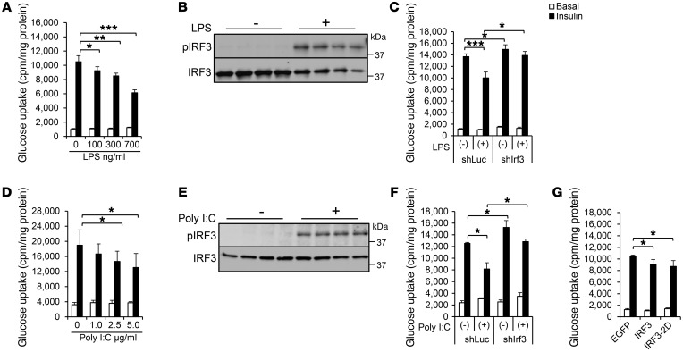
Figure 2. IRF3 regulates insulin sensitivity in adipocytes in a cell-autonomous fashion.
(A) Basal and insulin-stimulated glucose uptake in 3T3-L1 adipocytes after treatment with varying doses of LPS for 6 days. (B) Western blot showing phosphorylation of murine IRF3 (Ser388) in 3T3-L1 adipocytes after 6 days of LPS (700 ng/ml) treatment. (C) Basal and insulin-stimulated glucose uptake in 3T3-L1 adipocytes transduced with lentivirus expressing shRNA against Irf3 or shLuc control hairpin in the absence or presence of LPS treatment (700 ng/ml). (D) Basal and insulin-stimulated glucose uptake in 3T3-L1 adipocytes after treatment with varying doses of poly I:C. (E) Western blot showing phosphorylation of IRF3 in 3T3-L1 adipocytes after 24 hours of poly I:C (5 μg/ml) treatment. (F) Basal and insulin-stimulated glucose uptake in 3T3-L1 adipocytes transduced with lentivirus expressing shRNA against Irf3 or shLuc control hairpin in the absence or presence of poly I:C treatment (5 μg/ml). (G) Basal and insulin-stimulated glucose uptake in 3T3-L1 adipocytes expressing WT IRF3 or IRF3-2D mutant. Data in all panels expressed as mean ± SEM. *P < 0.05, **P < 0.01, ***P < 0.001.

