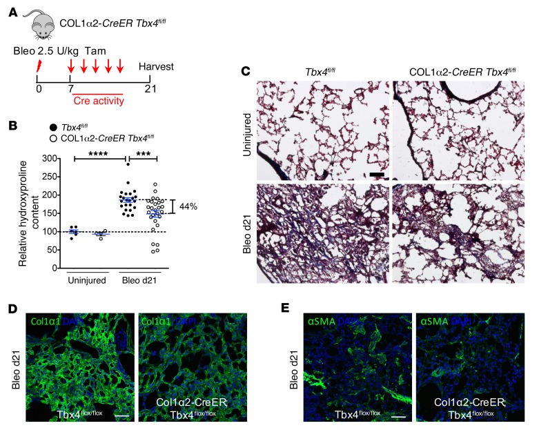
Figure 8. Loss of TBX4 expression in COL1α2-expressing cells attenuates pulmonary fibrosis.
(A) Strategy for inducible knockout of TBX4 expression in COL1α2+ cells. Col1a2-CreER Tbx4fl/fl mice were used in these experiments. The above-mentioned mice and their WT littermates (8–16 weeks old) were treated with bleomycin (2.5 U/kg), followed by 5 doses of tamoxifen (20 mg/g/injection) every other day starting on d7. The lungs were collected for hydroxyproline content determination on d21 after bleomycin. (B) Knockdown of Tbx4 in COL1α2-expressing cells decreased hydroxyproline content (means ± SEM, ***P ≤ 0.001, ****P ≤ 0.0001, n = 6 in uninjured Tbx4fl/fl group, n = 4 in uninjured Col1a2-CreER Tbx4fl/fl group, n = 23 in bleo Tbx4fl/fl group, n = 27 in bleo Col1a2-CreER Tbx4fl/fl group). (C) Representative Masson’s trichrome staining from lungs at d21 after bleomycin injection, showing decreased collagen deposition (blue) in Col1a2-CreER Tbx4fl/fl. (D and E) Representative images of COL1α1 and αSMA antibody staining for bleo d21 Col1a2-CreER Tbx4fl/fl mouse lung. n = 6 mice per group examined. Scale bars: 100 μm (C–E).

