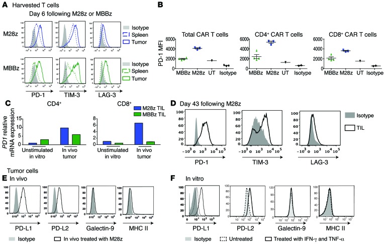
Figure 6. PD1 receptor and its ligands are upregulated in vivo.
(A) Tumor-infiltrating M28z and MBBz CAR T cells overexpressed inhibitory receptors 6 days after their administration. (B) MBBz CAR T cells express lower levels of PD-1 compared with M28z CAR T cells as shown by MFI of PD1 receptor expression of tumor-infiltrating CAR T cells (TIL) 6 days after intrapleural administration. Unstransduced tumor-infiltrating T cells (UT) express a low baseline level of PD1. (C) Relative expression of PD1 mRNA in CD4 and CD8 subsets of tumor-infiltrating CAR T cells 6 days after intrapleural administration. Data are represented as fold change relative to the PD1 mRNA expression of unstimulated M28z CAR+ T cells. (D) Tumor-infiltrating M28z CAR T cells isolated from progressing tumors express inhibitory receptors PD1, TIM-3, and LAG-3. (E) Single-cell tumor suspensions harvested from mice treated with M28z CAR T cells express high levels of PD-1–binding ligands. (F) In vitro–cultured mesothelioma tumor cells express the ligands (PD-L1, PD-L2) for the PD1 receptor, and expression is further upregulated following incubation for 24 hours with IFN-γ and TNF-α. Data are representative of at least 2 to 3 independent experiments.

