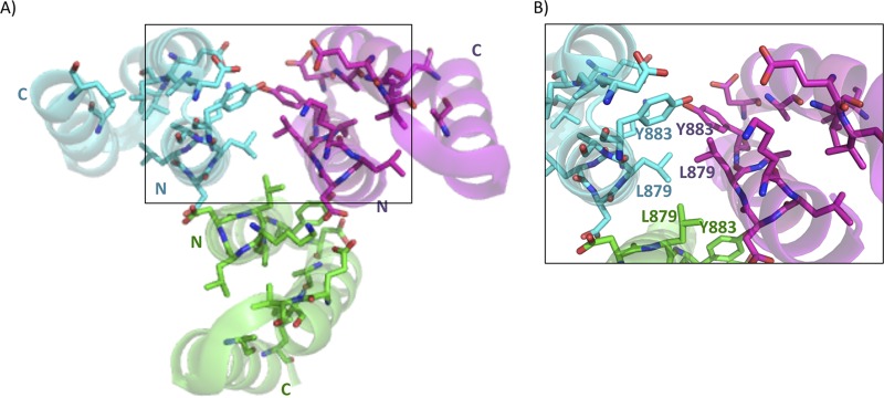
FIG 8.
X-ray crystal structure of FimV C-terminal domain homotrimer (PDB 4MBQ). The X-ray crystal structure of native FimV TPR3 was solved to 2.01 Å (PDB 4MBQ). Individual monomers are colored in cyan, green, or magenta. Highly conserved residues are shown as sticks with side chains colored by monomer. Residues and N and C termini are labeled in cyan, green, or magenta based on the associated monomer. Residues are numbered based on their position in full-length FimV. The boxed area is enlarged in panel B.

