FIG 3.

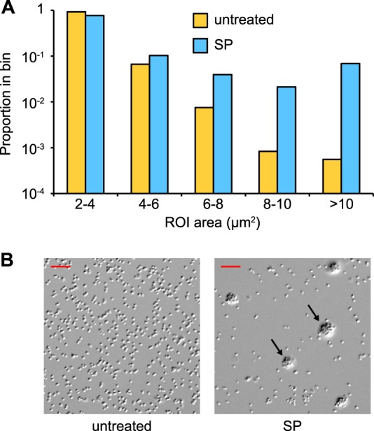
Exposure to SP stimulates formation of microcolonies. Suspensions of FA1090NV were incubated on glass coverslips for 1 h in the presence (SP) or absence (untreated) of 1:20 SP and imaged using DIC microscopy. (A) The distribution of ROI areas from 20 fields. The median ROI areas for untreated and SP-treated bacteria were 2.47 μm2 (n = 3,596) and 2.70 μm2 (n = 1,693), respectively. The distributions were significantly different by the Mann-Whitney rank sum test (U = 2,393,191; P < 0.0001). ROI areas of <2 μm2 were excluded from the analysis. (B) Representative DIC images from both conditions; arrows indicate microcolonies. Scale bars are 10 μm.
