Figure 5.
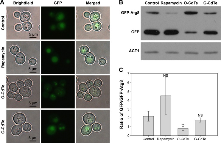
O-CdTe QDs induced accumulation of GFP-Atg8 in vesicles in yeast cells at 16 hours.
Notes: Cells with fluorescent tags were treated with 50 nmol/L CdTe QDs or 1 ng/mL rapamycin for 16 hours. (A) O-CdTe QDs partially inhibited the entrance of fluorescent dots into the vacuole and degradation. (B) Western blot analysis of intracellular GFP-Atg8 and GFP using antibodies to GFP. Actin (ACT1) was used as an internal reference. (C) Quantification of (B). The ratio of free GFP/GFP-Atg8 in each group was calculated. The results are expressed as mean ± standard deviation, n=3. **P<0.01, all versus controls.
Abbreviations: CdTe QDs, cadmium telluride quantum dots; G-CdTe, green-emitting CdTe; O-CdTe, orange-emitting CdTe; NS, no significance.

