
Figure 4.
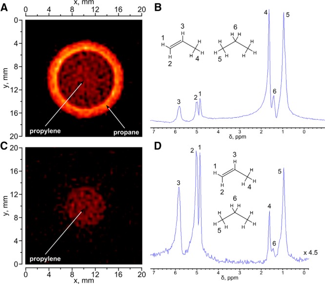
1H MR image (A) and corresponding 1H NMR spectrum of a sample comprising a 10-mm NMR tube with propylene placed inside a 15-mm tube with propane (B). 1H MR image (C) and corresponding 1H NMR spectrum (D) of the same phantom with application of selective suppression pulse for propane NMR signals. The FOV for both images was 5 × 5 cm with 128 × 128 matrix size and 10-mm slice thickness along the z axis; the total acquisition time was ∼8 seconds. The 1H NMR spectrum was obtained using the same ultrashort echo time (UTE) pulse sequence as that for 1H MRI but with the gradients switched off and the spectral bandwidth appropriately reduced.