Figure 2.
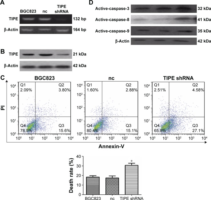
Suppression analysis of TIPE in the BGC823 cell line in vitro.
Notes: (A), (B) TIPE protein and mRNA levels expression in BGC823 cells that had been treated with shRNA were detected by Western blotting and RT-PCR. TIPE expression was suppressed by shRNA. (C) The rate of apoptosis for BGC823 treated with the Anti-DR5 ScFv was analyzed using flow cytometry, The apoptosis induced in the TIPE shRNA cells was enhanced compared with that induced in the negative control shRNA and the BGC823 cells. This image represents one of three sets of flow cytometry results. (D) The expression of the proteins involved in the apoptosis pathway of activated caspase-3/8/9. *P<0.05.
Abbreviations: nc, negative control; PI, propidium iodide.

