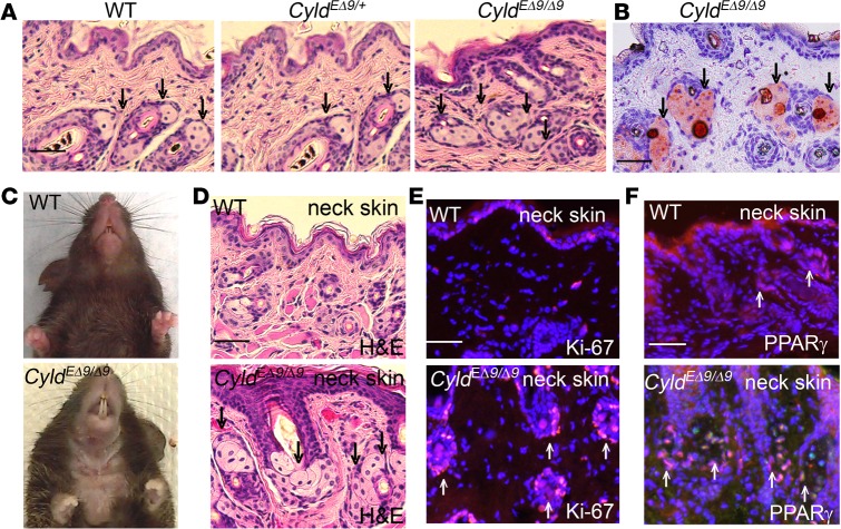
Figure 2. CyldEΔ9/Δ9 mice develop sebaceous gland hyperplasia.
(A) H&E staining of the back skin sections of 1-month-old WT, CyldEΔ9/+, and CyldEΔ9/Δ9 mice. (B) Oil Red O staining of the back skin sections of 1-month-old CyldEΔ9/Δ9 mice. (C) Clinical presentation of 5-month-old mice. (D) H&E staining of the neck skin of 5-month-old mice. (E–F) Immunostaining for Ki-67 and PPARγ (orange) of the neck skin of 5-month-old mice. Nuclei (blue, Hoechst 32558). Scale bars: 50 μm. Arrowheads mark sebaceous glands with negative staining by H&E and positive nuclei staining for Ki-67 and PPARγ.

