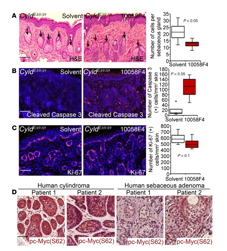
Figure 5. c-Myc is important for hair follicle–derived cell survival of CyldEΔ9/Δ9 mouse skin and is activated in human cylindroma and sebaceous adenoma.
(A) H&E staining of CyldEΔ9/Δ9 mice skin tissues treated with DMSO solvent or 10058-F4. Arrows mark sebaceous glands. Graph shows averages of sebaceous gland cells counted from 10 images of each condition. Data represent 25th–75th percentiles (box), median (line), and 5th and 95th percentiles (whiskers). (B–C) Immunostaining for cleaved caspase 3 and Ki-67 (orange); nuclei (blue, Hoechst 32558). Graphs show the average number of cells positively stained for cleaved caspase 3 and Ki-67 counted from 6 images of 2 different mice treated with the same condition. Data represent 25th–75th percentiles (box), median (line), and 5th and 95th percentiles (whiskers). P values were obtained via 2-tiered Student t test. (D) Immunoperoxidase staining of human cylindroma and sebaceous adenoma for phospho-c–Myc (S62) (brown) counterstained with hematoxylin. Two representative patient samples were shown for each group (n = 10). Scale bar: 100 μm.

