
Figure 5.
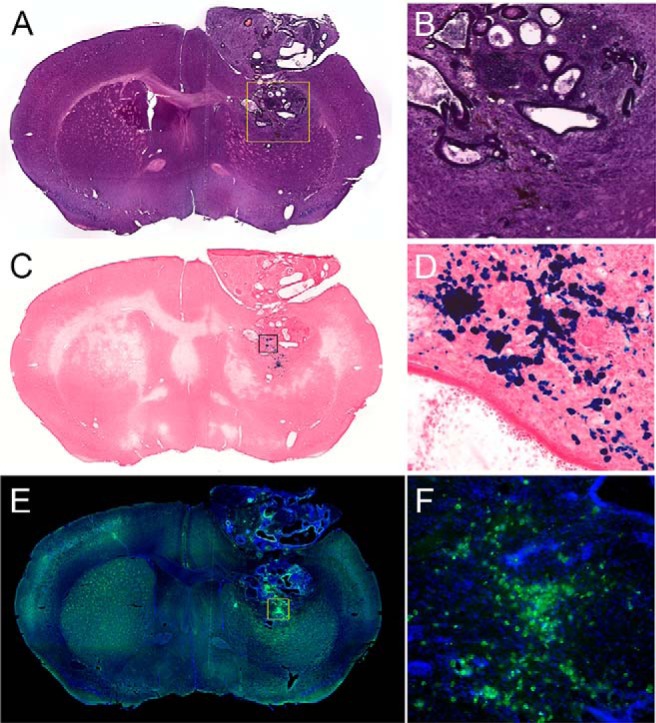
Hematoxylin and eosin (H&E)-stained coronal section showing tumor near implantation site (A, B). Prussian blue-stained section with nuclear fast red counterstain (C, D). Superparamagnetic iron oxide (SPIO) appears as blue deposits in the stain. Immunohistological stain for Luc (green) against DAPI (4′,6-diamidino-2-phenylindole) nuclear counterstain, showing Luc-expressing cells at both the original transplantation site and superficial lesion (E, F).