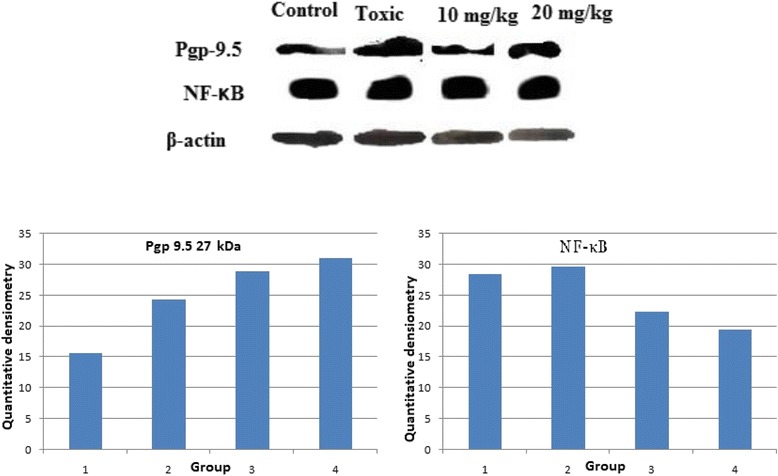
Fig. 4.

Western Blot analysis: Group I: Control; Group II: MNU; Group III: β-sitosterol10 mg/kg; Group IV: β-sitosterol 20 mg/kg. The western blot analysis containing whole tissue lysate from rat mammary gland was probed for P-gp 9.5 antibody; its immunoreactivity was evident as a band of molecular weight of 27 kDa. In case of control the expression is low in comparison with the toxicant. The low and high dose of β-sitosterol shows a similar pattern of elevated expression in a dose dependent manner. Results of the β-actin analysis are shown as an internal control
