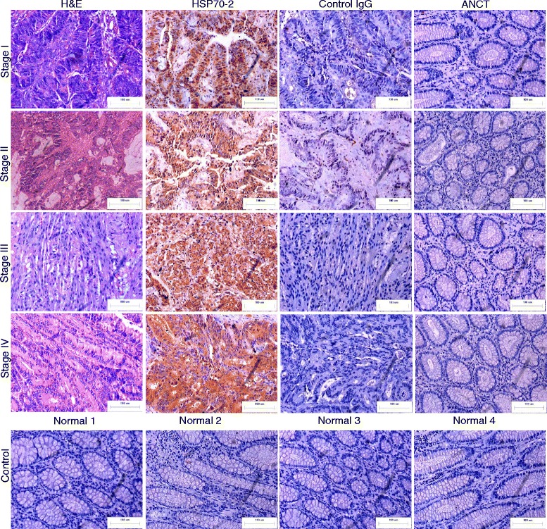
Fig. 2.

CRC specimens express HSP70-2 protein. First panel shows the cytostructure of representative micrographs of stage I-IV CRC specimen sections stained with H&E. Second panel shows chocolate brown reactivity in Stage I-IV CRC specimen sections probed with anti-HSP70-2 antibody. No immuno-reactivity was observed in stage I-IV CRC specimen sections probed with control IgG antibody (Third panel). ANCT specimens failed to show any immuno-reactivity probed with anti-HSP70-2 antibody (Fourth panel). Bottom most panel shows no HSP70-2 protein expression in representative specimens obtained from healthy patients. Original magnification x200, objective x20. Scale bar: 100 μm
