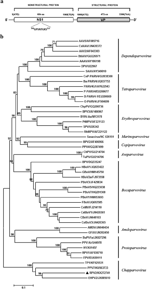
Fig. 1.

Genome organization and phylogenetic analysis of RPV2. a The genome rrganization of RPV2. The NS1 and VP proteins and the conservative ATP or GTP binding Walker loop motif are shown. b Phylogenetic tree was constructed over the NS1 amino acid sequences of 48 members in the Parvovirinae. The tree was derived using Neighbor-Joining analysis with 1000 bootstrap replicates. Scale bars indicate nucleotide substitutions per site, vertical bars represent the genera, and GenBank accession numbers are shown for the reference virus sequences, RPV2 identified in this study are marked with black triangle
