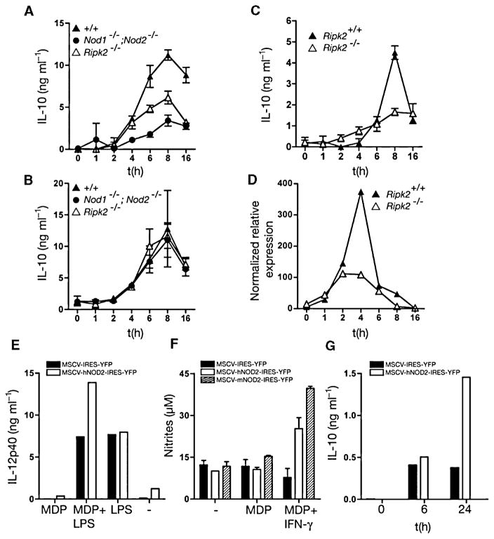
Fig. 4. IL-10 expression in response to PnCW is dependent on the RIPK2 pathway.
A. BMDMs from Ripk2−/− or Nod1−/−; Nod2−/− mice were stimulated with PnCW and IL-10 measured over time by ELISA.
B. As in (A), but following LPS stimulation.
C and D. Analysis of BMDMs from an independent Ripk2 mutant strain showing IL-10 and IL-10 mRNA produced in response to PnCW requires RIPK2.
E and F. Reconstitution of Nod2−/− hematopoietic stem cells with human or mouse NOD2 cDNAs rescues IL-10 production in response to PnCW. Stem cells from Nod2−/− mice were infected with retroviruses containing hNOD2 or mNOD2 cDNAs and selected by cell sorting for YFP and plated into media containing CSF-1 to drive differentiation into macrophages. After differentiation, macrophages were stimulated with LPS or LPS and MDP to measure the MDP synergy with TLR4 signalling (E) or IFN-γ signalling (F) or IL-10 production after PnCW treatment (G). Data are representation of two independent infection studies. In the case of mNOD2, sufficient cells were obtained to perform the MDP-IFN-γ synergy assay only.

