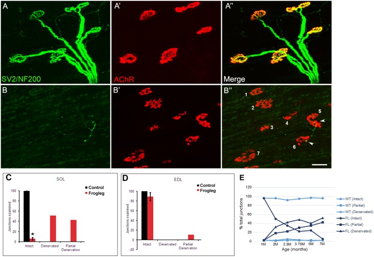
Fig 12. Immunolabeling of neuromuscular junctions of soleus and extensor digitorum longus (EDL) muscles.
Representative data for muscles isolated from 7 month old frogleg rat are shown in Panels A for the EDL and Panels B for the soleus. For the EDL, panel A shows staining (green) with antibodies to neurofilament (NF200) and synaptic vesicle protein (SV2), while panel A’ shows staining of the nicotinic acetylcholine receptors at the junction with α-bungarotoxin (red). A” shows the merged image, with yellow indicating normal innervation of each of the 5 junctions shown. In contrast, for the soleus, panel B indicates very little immunoreactivity with SV2 or NF200. Staining of the nicotinic acetylcholine receptors in B’ is normal, but in the merged image (B”) only 2 of the 7 numbered junctions show evidence of partial innervation (arrowheads at numbers 5 and 6), with the other 5 being fully denervated. Scale bar = 50 μm. Panels C and D show the cumulative data for soleus and EDL muscles respectively, for 7 month old animals. Black bars are for wild type and red bars for frogleg animals. In WT EDL and soleus muscles, 97.1% and 98.6% of NMJs are intact. In contrast, while the EDL NMJs were 89.8% intact in the frogleg mutant at 7M, only 4.4% of the NMJs for the soleus remained intact at that age. Signs of denervation were predominant in soleus muscles, with NMJs being either partially (42.6%) or completely (51.1%) denervated. Error bars = SD; * P< 0.05 (Relative to control). In panel E, the time course of denervation in the soleus muscle for frogleg and wild type rats aged 1–7 months is shown.

