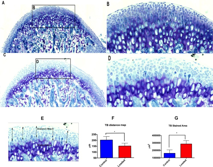
Fig 6. Altered loading increases proteoglycan accumulation.
The histological changes (TB Staining) within the mandibular condylar cartilage in the altered compressive loaded and control groups. Fig 6A–6B shows TB staining in the altered loaded group. Strong staining for proteoglycans is observed. The thickness of the mineralized cartilage is more. (6A) TB stained sagittal section of the MCC along with the subchondral bone, (6B) Intense proteoglycan staining in the fibrocartilaginous zone of the MCC, (6C-6D) TB staining in the altered unloaded group shows weaker staining when compared to the loaded group and less thickness of the mineralized cartilage. (6C) TB staining of the sagittal section of the MCC along with the subchondral bone in the unloaded group, (6D) thickness of mineralized fibrocartilage is less. (6F) Quantification of TB distance map, (6G) Quantification of TB stained area. Histograms represent means ± SD. * Statistically significant difference between the altered loaded side and the control group. Scale bar = 500μm.

