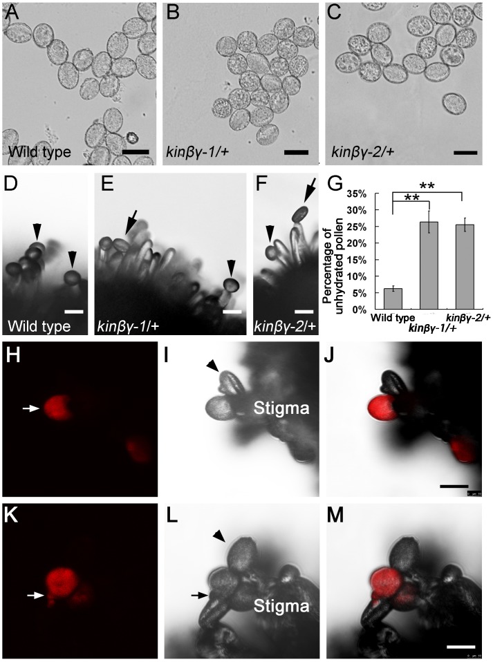
Fig 8. Pollen hydration in vitro and in vivo.
(A–C) Pollen of the wild type (A), kinβγ-1/+ (B) and kinβγ-2/+ (C) at 5 min after scattering onto the medium. (D-F) Pollen grains of the wild type (D), kinβγ-1/+ (E), and kinβγ-2/+ (F) at 5 min after pollinated on the wild-type stigmas. Arrowheads and arrows indicate hydrated and unhydrated pollen grains, respectively. (G) Statistical analysis of the number of unhydrated pollen grains of the wild type and kinβγ/+ at 5 min after pollinated on the wild-type stigmas. Data were collected from three independent experiments. Asterisks indicate significant difference (Student’s t-test, P<0.01). (H–J) Hydration of the kinβγ-2/+ dsred/+ qrt1/- pollen grains on the wild-type stigmas at 5 min after hand-pollination. Arrow in (H) and arrowhead in (I) indicate a hydrated (red fluorescence signal) and an unhydrated (no red fluorescence signal) pollen grains in a tetrad, respectively. (H–J) The photographs of fluorescence channel, bright-field channel, and the merged channel, respectively. (K–M) Germination of the kinβγ-2/+ dsred/+ qrt1/- pollen grains on the wild-type stigmas at 25 min after hand-pollination. Arrows in (K, L) and arrowhead in (L) indicate a pollen tube with red fluorescence signal and a hydrating pollen grain without red signal, respectively. (K), (L), and (M) The photograph of fluorescence channel, bright-field photograph, and the merged photograph, respectively. No mounting medium was used in the preparation of the temporary slides for the observations and photographs of (D–F, H–M). Bars, 20 μm.

