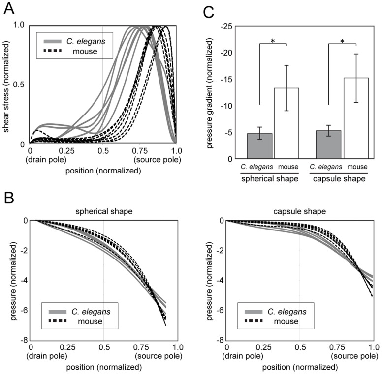
Fig 6. Shear-stress distribution in mouse oocytes contributes to the generation of a pressure gradient that enables the positioning of the meiosis II spindle near the cell surface.

(A) Comparison of shear-stress distributions of the C. elegans embryo and mouse oocyte showing that shear stress is localized closer to the cell periphery in the latter. (B) Pressure when flow is generated using the estimated shear stress plotted against source-drain position. The plot shows that the gradient is steeper when we assume the shear-stress distribution in the mouse oocyte rather than that in the C. elegans embryo in both spherical- and capsule-shaped cells. (C) Comparison of the pressure gradient at the source end in Fig 6B; the pressure gradient is steeper in the mouse oocyte than in the C. elegans embryo. *P < 0.005 (t test, assuming non-equal variance).
