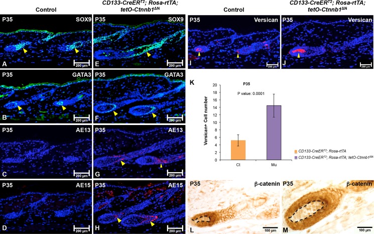
Fig 4. Expression of ΔN-β-catenin in CD133+ DP cells accelerates hair follicle differentiation.
5-μm-thick paraffin sections from P35 CD133-CreERT2; Rosa-rtTA; tetO-Ctnnb1ΔN mutant mice and control littermates were processed for immunofluorescence staining of following markers: Sox9 for outer root sheath (control: A; mutant: E); Gata3 for inner root sheath (control: B; mutant: F); AE13 and AE15 for hair keratins (control: C, D; mutant: G, H); versican for anagen DP (control: I; mutant: J). Sections were nuclear counterstained with DAPI (blue). K. The numbers of versican+ DP cells in each hair follicle were counted and compared between CD133-CreERT2; Rosa-rtTA; tetO-Ctnnb1ΔN mutant mice and control littermates (mean ± s.d). A minimum of three skin biopsies from three pairs of mutant and control mice was analyzed. Two-tailed paired Student’s t-test was employed to calculate statistical significance. (L-M) β-catenin expression in hair follicle was examined by immunohistochemistry. Images shown are representative of at least three replicates at each indicated age. Scale bars: 200 μm for A-J; 100 μm for L-M.

