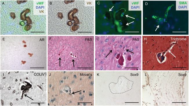
Figure 2.

BGC is localized to cerebral arterioles. The calcified lesions (arrows in C) were localized to arterioles, characterized by the co‐localization of vWF and von Kossa (VK) staining and counterstaining with the nuclear marker DAPI (A‐C). Furthermore, the lesions (arrow in D) were also present in SMA‐positive vessels counterstained with DAPI (D). Alizarin Red (AR) staining compared with Periodic acid‐Schiff (PAS)‐positive staining of consecutive sections confirmed that the calcified lesions (arrows in F) contained basement membrane components (E‐G) and localized to blood vessels (arrows in G). Positive Masson's Trichrome staining indicated that the lesions were rich in collagen and/or mucin (H) and formed in blood vessels (arrow in Figure H). Immunohistochemical staining identified collagen type IV as the major basement membrane protein present in the calcified lesions (I) and further confirmed localization to blood vessels (arrow in I). The SMC in the calcified regions do not undergo an osteochondrogenic phenotype change, as the lesions (arrow in J) lacked proteoglycan‐rich or cartilage‐like structures, confirmed by negative Movat Pentachrome (J) and SOX9 (K) staining. Calcified aortic tissue from a diabetes mouse model was used as a positive control (L). Scale bars: 50μM (A‐D,G‐L), 200μM (E,F).
