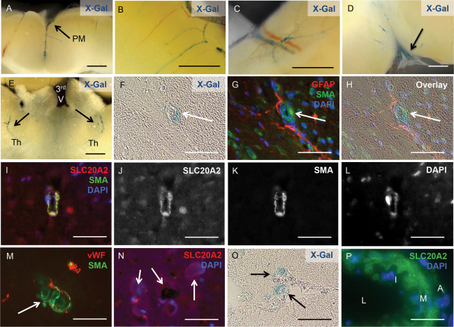
Figure 6.

Slc20a2 is highly expressed in cerebral SMC. X‐Gal activity was detected in arteries within the pia mater (PM) (A), the cortex (B), the cerebellum (C), in the region of the cervical lymphatics (D), and in the thalamus of both hemispheres (Th); location of the third ventricle (3rd V) is indicated for spatial orientation (E). X‐Gal staining and anti‐SLC20A2 antibody signal co‐localized with SMA‐positive cells and GFAP‐positive cell projections (F‐L). Calcified lesions (arrows in M, N) also co‐localized with vWF and SMA positive cells (M), and revealed the presence of SLC20A2 positive cells in low numbers (N). Slc20a2 was also expressed in media of vessels that connect to the CP (O) and in large cerebral arteries (L: lumen, I: intima, M: media, A: adventitia) (P). Scale bars: 2mm (A‐E), 50μM (F‐P).
