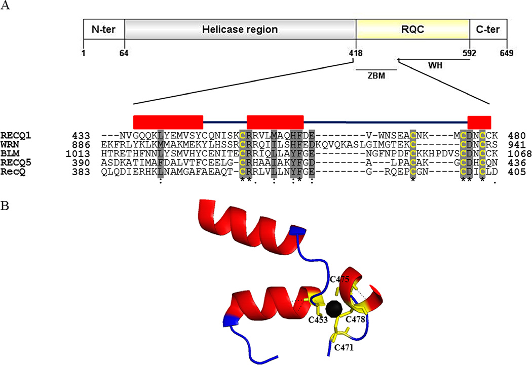
Figure 1. Schematic representation of RECQ1 protein.
(A) Conserved domains in RECQ1 and amino acid sequence alignment of the conserved zinc binding domain (ZBD) among the RecQ family helicases in humans. The multiple sequence alignments were generated with MUSCLE and refined manually. Positions of the first and the last amino acid residues are shown by the numbers at the beginning and end of each sequence, respectively. Highly conserved residues are shadowed in gray color. In bold yellow color are the four conserved cysteine residues. Secondary structure elements of the RECQ1 are shown with boxes designating the α-helices. (B) Ribbon drawing of the ZBD of the crystallized truncated RECQ1 protein (PDB ID: 4U7D). The zinc ion shown as a black sphere is coordinated by four conserved cysteine residues.

