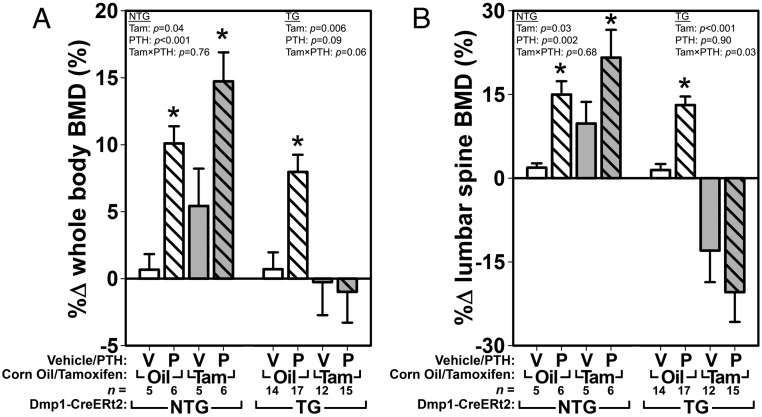
Figure 2.
DEXA-derived measures of BMD using whole-body (A) and lumber spine (B) ROIs, calculated as the percent difference from 12 weeks of age (beginning of Cre induction and PTH treatment) to 17 weeks of age (end of PTH treatment), reveal that PTH treatment failed to increase BMD when βcat was deleted from 10kbDmp1-expressing cells. The left side of each graph indicates the effect of tamoxifen (Tam) and PTH (P) in essentially wild-type mice (no Cre transgene present [NTG] to recombine the floxed βcat alleles that are present in all groups). The right side of each graph indicates the effect of PTH in TG mice with (Tam) or without (Oil) βcat deletion. Among the TG mice, whole-body BMD and spine BMD ANOVAs (upper corners) yielded near significant (P = .06) and significant (P = .03) interaction terms, respectively, suggesting that βcat deletion significantly affects the efficacy of PTH (at least in the spine). Asterisks indicate significant (P < .05) PTH-induced difference from the tam/oil-matched vehicle control, using PLSD post hoc pairwise tests.

