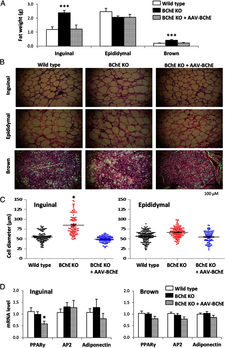
Figure 5.
Adipose tissue weight, adipocyte cell size, and adiposity-related gene expression. A, Inguinal sc, epididymal, and brown adipose tissue depot weights in 10-month-old controls, BChE KO, and vector-rescued BChE KO mice on HFD. B, H&E-stained sections of adipose tissues. Scale bar, 100 μm. C, Cell diameters of isolated inguinal and epididymal fat pads. D, mRNA levels of adipogenesis markers (PPARγ, adipocyte protein 2 [AP2], and adiponectin). Results are mean ± SEM (n = 9–10); *, P < .05; ***, P < .001, relative to wild-type control.

