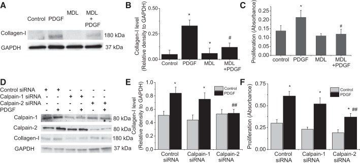
Fig. 2.
Inhibition of calpain-2 prevents PDGF-induced collagen synthesis and proliferation of PASMCs. A–C: PASMCs were incubated with PDGF-BB (10 ng/ml) in the absence and presence of MDL28170 (MDL; 20 μM) for 24 h, after which collagen I and cell proliferation were measured. D–F: PASMCs were transfected with calpain-1 or calpain-2 siRNA or control siRNA and then incubated with PDGF-BB (10 ng/ml) for 24 h, after which collagen I and cell proliferation were measured. A and D: representative immunoblots of collagen I. B and D: bar graphs showing the changes in protein levels of collagen I. C and F: bar graphs showing the changes in cell proliferation. Values are means ± SE; n = 4. *P < 0.05 vs. control. #P < 0.05 vs. PDGF only. ##P < 0.05 vs. control siRNA + PDGF.

