Fig. 6.
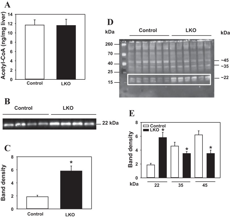
Hepatic acetyl-CoA (AcCoA) content and analyses of acetylated proteins in liver nuclei from L-PDCCT (control) and L-PDCKO (LKO) mice. A: hepatic AcCoA content in mice. B: Western blot analysis of histone H3 acetyl K9 protein (H3K9) in liver nuclei from mice. C: densitometry analysis of histone H3 acetyl K9 protein detected in B. D: Western blot analysis of acetyl-lysine proteins in liver nuclei. E: densitometry analysis of 22-, 35-, and 45-kDa acetyl-lysine proteins bands detected in D. Protein loading was analyzed by staining of gels with Ponceau S stain, followed by densitometry analysis (results not shown). Results are expressed as means ± SE (n = 6–8). *P < 0.05.

