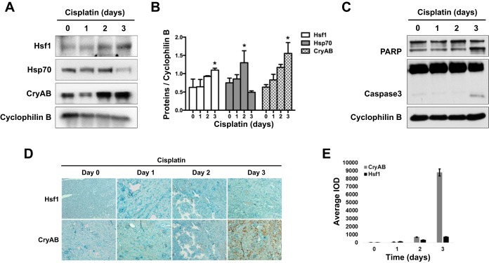
Fig. 1.
Activation of the heat shock response in cisplatin-induced nephrotoxicity in mice. B6 mice were given 30 mg/kg cisplatin via one intraperitoneal injection. Animals were then euthanized at days 1, 2, and 3 after the injection. A: immunoblot analysis of the heat shock response in kidney tissues during cisplatin treatment of mice. Kidney cortical tissue lysate was collected for immunoblot analysis of heat shock factor (Hsf)1, heat shock protein (Hsp)70, crystallin-αB (CryAB), and cyclophilin B (internal loading control). B: quantification and statistical analysis of Hsf1, Hsp70, and CryAB proteins were performed using ImageJ software and two-way ANOVA. Data are expressed as means ± SD; n = 3. *P < 0.05 vs. the other three time points of cisplatin treatment. C: immunoblot analysis of apoptotic poly(ADP-ribose) polymerase (PARP) and caspase 3 cleavage. Kidney tissue lysate was subjected to immunoblot analysis of PARP, caspase 3, and cyclophilin B. D: immunohistochemical staining of Hsf1 and CryAB in kidney tissues. Kidney tissue sections were subjected to immunohistochemical staining using a streptavidin-biotin system and the antibodies specific to anti-CryAB and anti-Hsf1 antibodies. E: quantification of the immunohistochemical staining of Hsf1 and CryAB in kidney tissues. The average integrated optical density (IOD) value of each slide was calculated using Image-Pro plus 6.0 software.

