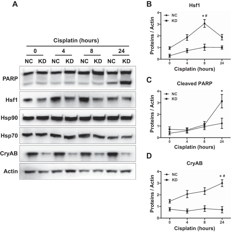
Fig. 5.
Hsf1 KD lead to a downregulation of CryAB in RPTCs. Hsf1 KD cells and NC cells were incubated with 20 μM cisplatin for 0, 4, 8, and 24 h. A: whole cell lysates were collected for immunoblot analysis of Hsf1, PARP, CryAB, Hsp90, and Hsp70. The same blots were reprobed for β-actin. B–D: quantification of Hsf1 (B), cleaved PARP (C), and CryAB (D) proteins was performed using ImageJ software. Data are expressed as means ± SD; n = 3. *P < 0.05 vs. KD cells treated with cisplatin at the same time point (Student's t-test); #P < 0.05 vs. NC at the other three time points of cisplatin treatment (two-way ANOVA).

