Fig. 1.
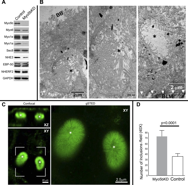
Myo5bKD C2BBe cells model MVID villus enterocytes. A: immunoblots of Myo5b, Myo6, Myo1e, Myo1a, Sec 8, NHE3, EBP50, NHERF2, and GAPDH (for protein load) from equivalent protein loads of lysates prepared from fully polarized mature control (scrambled) and Myo5bKD C2BBe cells. B: transmission electron micrographs (TEM) of mature Myo5bKD C2BBe cells show apical microvillus inclusion (MVI) with brush border (black asterisk) at low-power (left, scale bar 2 μm) and high-power (middle, scale bar 800 nm) magnification and basolateral MVI with BB (black asterisk) at low-power (right, scale bar 2 μm) magnification. BB, brush border; BLM, basolateral membrane. C: confocal (XZ and XY, scale bar 20 μm) and gSTED images of a bracketed XY confocal area shows actin labeling in green. Asterisk marks MVIs. Scale bar, 2.5 μm. D: quantification in fully polarized Transwell-grown C2BBe cells reveals increased number of inclusions in Myo5bKD C2BBe cells (expressed as a total number per ×40 power field of view).

