Fig. 5.
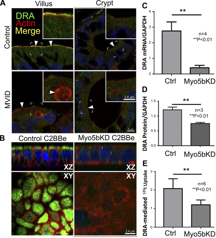
DRA localization, expression, and function in the human duodenum, control, and Myo5bKD C2BBe cells. A: confocal microscopy images of DRA (green) IFL in cryosections from normal control and MVID human duodenum are shown in relation to F-actin (red); nuclei are stained in blue. Brackets circumscribe the areas inserted under higher magnification, arrowheads indicate location of DRA label in merge images. Inset, scale bar 2.5 μm; main panel, scale bar 5 μm. B: confocal Z-stack XY and XZ projections of Transwell-grown C2BBe monolayer cells. DRA (green) staining is shown in relation to F-actin (red); nuclei are stained in blue. Scale bar, 2.4 μm. C: DRA mRNA levels relative to GAPDH in mature polarized C2BBe cells. D: quantification of DRA protein relative to GAPDH in lysates of mature polarized C2BBe cells. E: Cl−/HCO3− exchange using DIDS-sensitive 125I uptake in mature polarized C2BBe cells.

