Fig. 8.
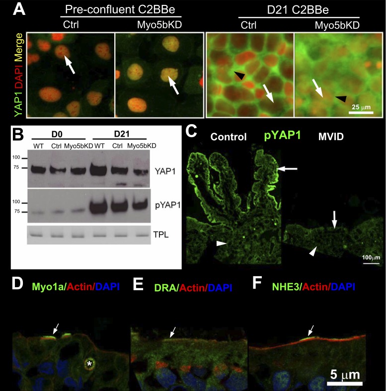
YAP1 localization, expression, and phosphorylation in preconfluent and mature polarized day 21 C2BBe cells. IFL of pYAP1 distribution in normal and MVID human intestine and BB enterocyte compensatory responses in MVID. A: confocal microscopy images of YAP1 (green) and nuclear DAPI (red, arrow) IFL in control and Myo5bKD preconfluent (D0) and mature polarized (D21) C2BBe cells. Black arrowhead indicates cytosolic label. Scale bar, 25 μm. B: immunoblots of YAP1 and phospho-YAP1 (pYAP1) in lysates of prepolarized day 0 (D0) and polarized day 21 (D21) C2BBe cells is shown relative to the total protein load (TPL). C: IFL of pYAP1 (green) in crypt (arrowhead) and villus (arrow) of normal (left) and MVID (right) human intestine. Scale bar, 100 μm. D–F: confocal images of IFL of BB proteins in villus sections of human MVID duodenum. F-actin is shown in red and nuclei in blue in all panels. D: Myosin1a (green) labels the BB in two MVID enterocytes (arrows) and decorates an MVI marked with an asterisk. E: DRA (green) IFL in the BB of cells marked (arrow) is higher than in its neighbors. F: NHE3 (green) IFL in the BB of some enterocytes is significantly higher (arrow) than neighboring cells. Scale bar, 5 μm.

