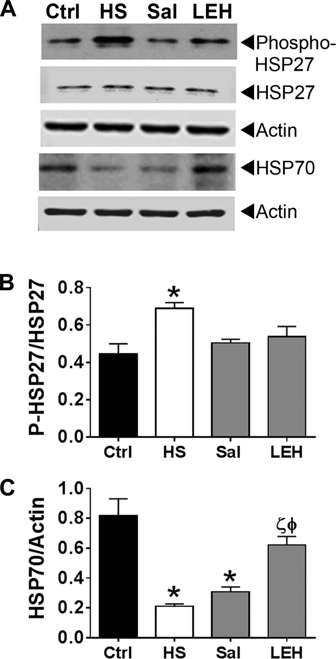
Fig. 3.
Expression of heat shock proteins in hemorrhagic shock and resuscitation. A: representative immunoblot showing the expression of phospho-HSP27 and HSP70. Actin was used as a loading control. B and C: densitometry of the HSP27 and HSP70 immunoblots, respectively (n = 3 per group). The immunoreactive band density was normalized with the density of actin band. P < 0.05 vs. Ctrl*, HSϕ, and Salζ. Additional immunoblots of phospho-HSP27 and HSP70 are provided in the supporting information.

