Fig. 9.
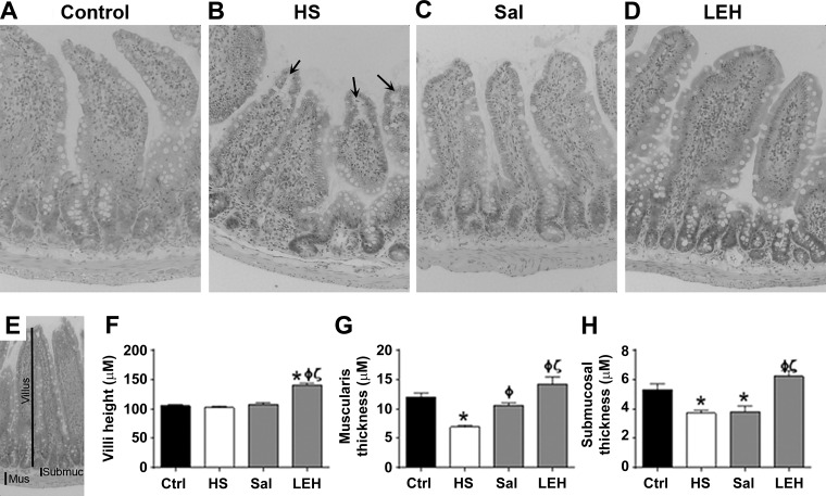
Histopathology. Control (A), HS (B), saline (C), and LEH (D). Arrows in B point to the subepithelial spaces observed under the microscope. E: depiction of morphometry performed on light microscopic pictures; the thickness of muscularis (mus), submucosa (submuc), and villus are shown. Villus height (F), muscularis thickness (G), and submucosal thickness (H).

