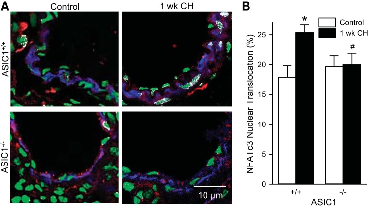
Fig. 2.
ASIC1 contributes to CH-induced NFATc3 nuclear accumulation. Representative images (A) and summary data (B) showing NFATc3 nuclear translocation (white) was assessed by % NFATc3 colocalization (red) with SYTOX nuclear stain (green) in smooth muscle α-actin-positive cells (blue) in fixed lung sections from control and CH-exposed (1 wk) ASIC1+/+ and ASIC1−/− mice. Data are expressed as means ± SE, n = 32 arteries from 4 animals per group. *P < 0.05 vs. control mice, #P < 0.05 vs. ASIC1+/+ mice analyzed by a two-way ANOVA and individual groups compared with the Student-Newman-Keuls test.

