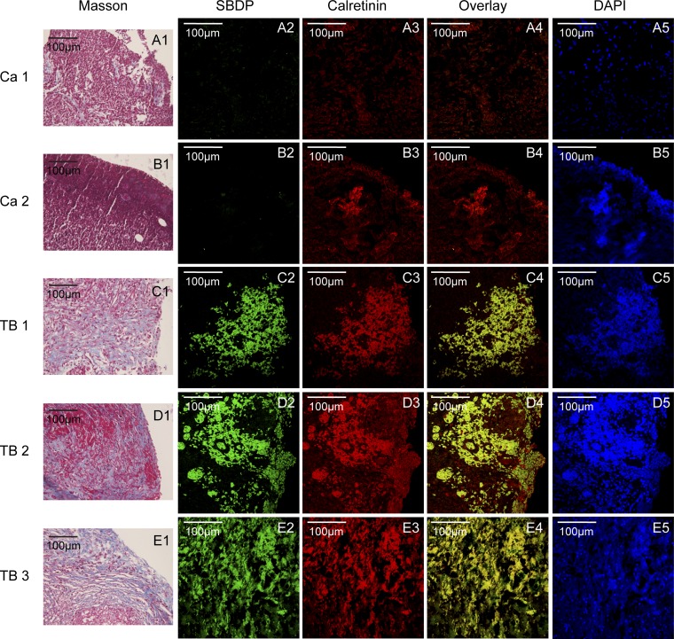
Fig. 7.
Activation of calpain and collagen deposition in the pleural samples from patients with tuberculous pleurisy. Pleural tissue samples were collected from parietal pleura in patients with TPE (TB 1, 2, 3) or MPE (Ca 1, 2). The pleural sections were stained with Masson staining for morphological analysis. spectrin breakdown product (SBDP) and calretinin (marker for PMCs) were stained by immuofluorescence staining. A1–E1: Masson stainings, blue color shows collagen. A2–E2: immuofluorescence stainings of SBDP (green color). A3–E3: immuofluorescence stainings of calretinin (red color). A4–E4: overlays of immuofluorescence stainings, yellow color shows colocalization of SBDP and calretinin. A5–E5: DAPI stainings.

