Fig. 2.
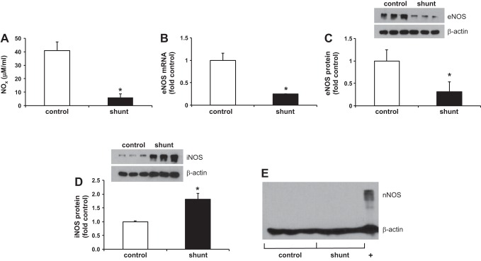
Bioavailable NO (NOx) and endothelial nitric oxide synthase (eNOS) expression is decreased in shunt LECs, but inducible nitric oxide synthase (iNOS) expression is increased. A: NOx levels in control (40.9 ± 6.3 μM) and shunt (6.1 ± 2.7 μM) LECs, *P < 0.001. B: eNOS mRNA expression is decreased 4-fold in shunt LECs, *P < 0.05. Relative RNA expression quantified by quantitative real-time PCR (qPCR) and normalized to control. Data are shown as means ± SE. C: eNOS protein expression is decreased 3-fold in shunt LECs, *P < 0.05. D: iNOS protein expression is increased 1.8-fold in shunt LECs, *P < 0.01. E: neuronal nitric oxide synthase (nNOS) is not expressed in control or shunt LECs; +, positive control (tissue homogenate from right ventricle) for nNOS. Protein levels in control and shunt LECs quantified by Western blot. For presentation graphically, densitometry in each lane has been normalized to β-actin and to control. For all experiments, n = 3 control, 3 shunt.

