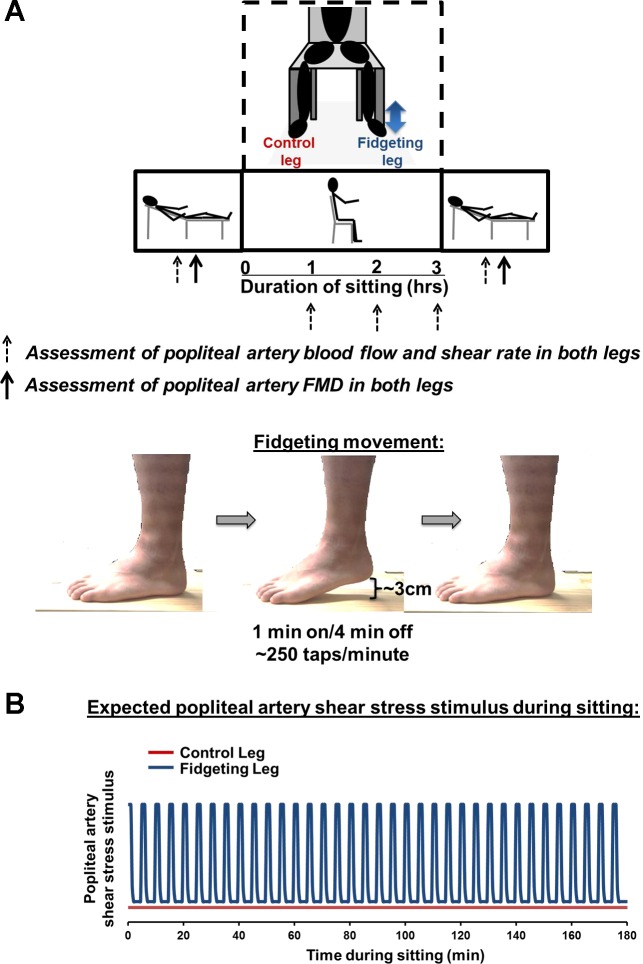
Fig. 1.
Experimental design. A: schematic diagram of experimental protocol and positional changes over the course of the study. Measurements taken at the time points of 1, 2, and 3 h were made while the subject was in the seated position, whereas presit and postsit measurements were taken while subject was in the supine position. B: expected popliteal artery shear stress stimulus during sitting in the control and fidgeting legs. FMD, flow-mediated dilation.

